Dr. Nadeem Murtaza, (L), is a postdoctoral researcher in the lab of Dr. Karun Singh, Senior Scientist at the Donald K. Johnson Eye Institute and the senior author of the study. (Photo: UHN StRIDe Team)
Researchers at University Health Network’s Donald K. Johnson Eye Institute have mapped protein networks in brain cells to clarify how genes associated with autism spectrum disorder interact with one another.
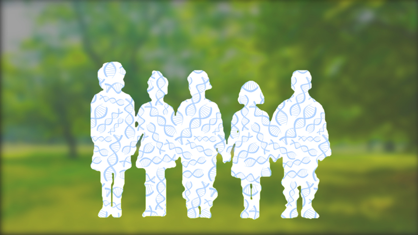
Autism affects one in 50 Canadian children and youth and there is no cure. Individuals with this neurodevelopmental disorder often experience difficulties with learning, communication and social interactions.
Researchers have identified hundreds of risk genes for autism, but it is still unclear how genetic changes affect downstream cellular processes to trigger the condition. Because genes code for proteins, exploring how genetic mutations translate into changes in protein activity could provide deep insights into the root causes of autism and how to treat it.
“A long-standing question in autism research is whether risk genes act alone or through shared signalling pathways to cause the disorder,” says Dr. Karun Singh, a Senior Scientist at Donald K. Johnson Eye Institute and the senior author of the study.
“A unique element of this study is that we used proteomics – the large-scale study of the structure and function of proteins – to determine if and how risk genes converge onto shared molecular pathways in neurons,” adds Dr. Singh, who is also an associate professor in the Department of Ophthalmology & Vision Sciences at the University of Toronto.
Dr. Singh’s team developed a protein-mapping technique to identify interaction networks for the proteins that are encoded by 41 known autism risk genes.
The team discovered that many of the proteins encoded by these genes play important roles in basic neuron functions, such as cell-to-cell communication and energy production. While these findings highlight the known role of abnormal neuron communication in autism, they also point to a less well-studied disease mechanism: changes in cellular metabolism.
Using gene-editing techniques, the team confirmed that several of the risk genes regulate the activity of mitochondria, the energy factories within cells. Because brain cells are metabolically very active, disruptions in their mitochondrial activity can dramatically disrupt brain function.
The link between risk genes and mitochondrial dysfunction sheds light on how these genes might change brain cell activity and ultimately cause autism symptoms.
The next step for this research is to apply the protein-mapping technique to patient-specific brain tissues that the team generates from stem cells created from patients’ blood. These tissues display the patients’ unique gene and protein profiles and can be used to identify molecular signatures of their disease.
“Protein screening has the potential to reveal disease mechanisms that we can use to classify patients based on underlying biological processes, rather than symptoms alone,” says Dr. Nadeem Murtaza, a postdoctoral researcher in Dr. Singh’s lab. “Because autism is a highly variable disorder, identifying objective ways to classify patients with the condition is an important next step towards unlocking more personalized treatments.”
This work was supported in part by donors to UHN Foundation.