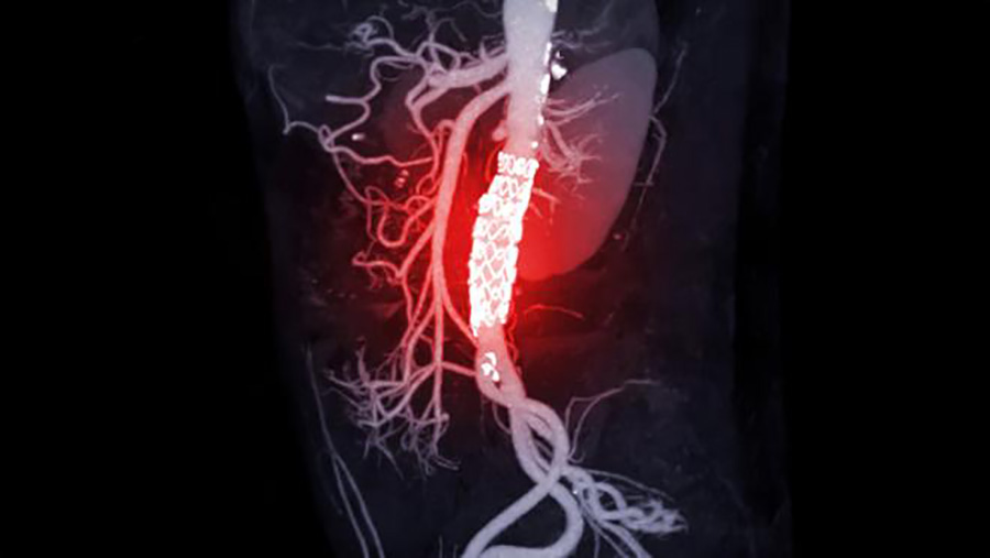
Researchers at UHN’s Toronto General Hospital Research Institute (TGHRI) have utilized artificial intelligence (AI) to identify safe and dangerous zones for deploying a stent graft – a tube made of a metal with a fabric covering that protects the aorta – during surgery to repair an abdominal aortic aneurysm, a condition where the aorta in the abdomen becomes enlarged and weak.
The aorta is the main artery that carries blood from the heart to the rest of the body. An abdominal aortic aneurysm (AAA) can rupture, which is a life-threatening complication.
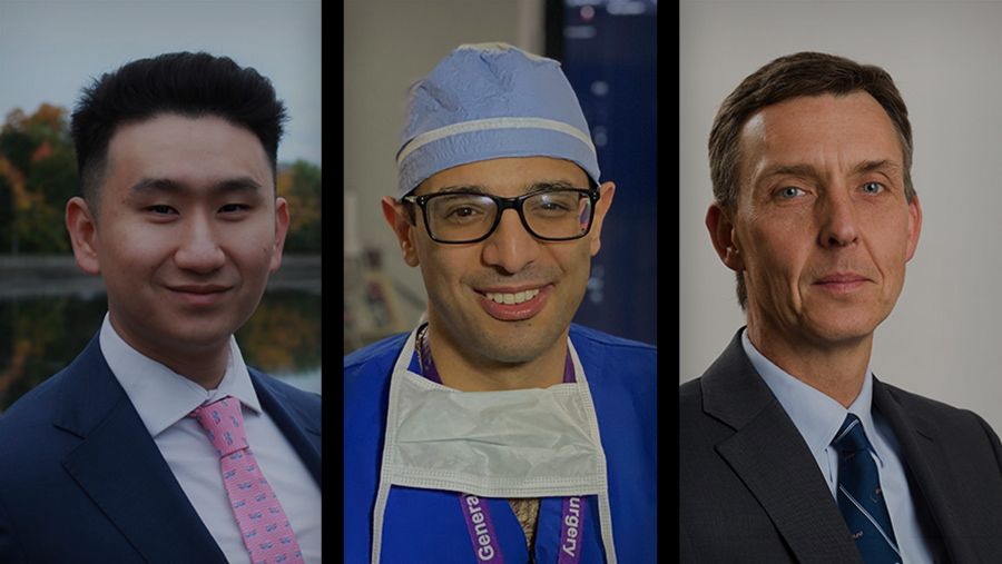
“Endovascular aneurysm repair (EVAR) is a common method of treatment for AAAs where a stent graft is inserted to act as a lining for the aorta and lower the risk of a rupture,” says Dr. Thomas Forbes, UHN’s Surgeon-in-Chief, a clinician investigator at TGHRI and senior author of the study.
“Current practice for this procedure involves manually identifying the correct area of the aorta using fluoroscopy – videos of movement inside the body through the use of X-rays.”
A dangerous complication of this surgery is the partial or complete blockage of blood flow in the renal artery by the deployed stent graft. This can occur in part due to errors in visual perception and judgement when positioning the stent.
To try and prevent this complication, TGHRI researchers developed a deep learning model trained to identify “no-go’ landing zones – areas where insertion of the stent graft would cause a blockage – during EVAR surgery.
“We used 369 fluoroscopic images from 110 different patients/videos to train our AI model,” says Allen Li, medical student at the University of Ottawa and first author of the study. “These images were obtained from previous EVAR procedures performed at a single institution (UHN) and from open-access sources.”
Dr. Amin Madani, co-author of the study, endocrine surgeon at UHN and Director of the Surgical AI Research Academy (SARA) says: “Our results showed that AI was able to successfully identify the area of the abdominal aorta during surgery where insertion of the stent graft would cause renal blockage – the ‘no-go’ zone.
“However, more work is needed to optimize the AI model’s performance when using datasets from more than one institution.”
Dr. Forbes, who is also a professor and Vice-Chair in the Department of Surgery at U of T, adds: “This study shows that AI can be used as an intraoperative tool and methods from this study may be useful for other types of endovascular surgeries.
“These results provide further insight into the use of AI in surgery applications as it becomes an increasingly important part of the operating room.”
This study was supported by generous donors to UHN Foundation.
No one ever changed the world on their own but when the bright minds at UHN work together with donors we can redefine the world of health care together.