Imagine this: you’re having a heart attack. Almost immediately, scar tissue begins to form around your heart, and it becomes permanently damaged. But what if, when you arrived at the hospital, a doctor could treat you with a drug that helped your heart generate new cells and rebuild itself?
Or, imagine you have breast cancer and you’re undergoing long rounds of chemo and radiation therapy, treatments that could harm your heart. Besides monitoring the cancer, your cardiologist could also detect signs of heart trouble early, adjust your chemo regimen — without inhibiting its success — while preventing you from getting heart disease, which is currently the leading cause of death in breast cancer survivors.
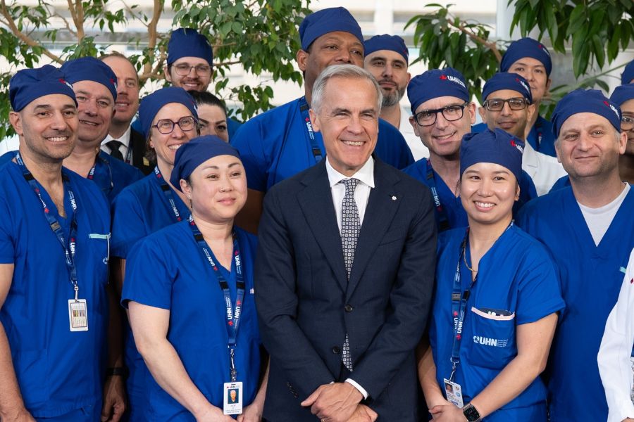
Researchers are doing all this and more, pioneering cutting-edge approaches to healing damaged hearts, increasing the number of hearts available for transplant and preventing the risk of stroke. Dr. Kathryn Howe, a vascular surgeon at the Peter Munk Cardiac Centre, Division of Vascular Surgery, UHN Sprott Department of Surgery, says every new research finding is driven by the one question physicians ask themselves daily: “How can we have something better to offer?”
Here’s some of the groundbreaking work underway at the Peter Munk Cardiac Centre.
The troubleshooter
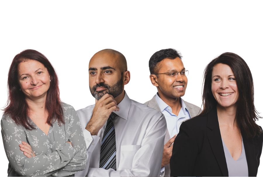
Who: Dr. Paaladinesh Thavendiranathan, Director, Ted Rogers Cardiotoxicity Prevention Program, Ted Rogers Centre for Heart Research, Peter Munk Cardiac Centre and a cardiologist at the Centre
The goal: Preventing heart damage for patients undergoing cancer treatment.
The research: After having undergone intense radiation or chemotherapy, cancer survivors shouldn’t be faced with the burden of a second life-threatening illness, yet unfortunately many can develop heart disease. “Survivors can be left with arrhythmias [irregular beating of the heart], heart muscle dysfunction and heart fibrosis, all of which can lead to heart failure,” Dr. Thavendiranathan explains.
He’s currently studying how the heart is affected in women with breast cancer. Traditionally, assessing a patient’s heart is done with an echocardiogram (a photo of the heart) that measures the percentage of blood leaving the heart each time it pumps. But, he says, “at that point, we’re too late.”
Using ultrasound and magnetic resonance imaging (MRI), patients are assessed prior to treatment and every three months thereafter to see if there are early indicators of heart inflammation or fibrosis. “I’m going to pick up on heart dysfunction before anyone else does,” says Dr. Thavendiranathan. If he and his team can catch changes early on, patients can have their chemotherapy tweaked or be given heart-protective medications.
The sleuth
Who: Dr. Kathryn Howe, vascular surgeon, Peter Munk Cardiac Centre, Division of Vascular Surgery, UHN Sprott Department of Surgery
The goal: Preventing strokes by identifying which arterial plaques are dangerous.
The research: Buildup of plaque in the carotid artery can be inactive — or it can be deadly. Unfortunately, physicians can’t easily tell which arterial plaques are problematic. “Some of the plaques are stable for a lifetime,” says Dr. Howe.
In other cases, increased inflammation causes the plaque to become unstable, which can potentially result in necrosis (cell death). Plaque can also break off and form a blood clot, leading to a stroke.
Dr. Howe is analyzing how artery-lining endothelial cells use extracellular vesicles (particles released from a cell) to communicate with plaque cells to form plaques. The contents of some extracellular vesicles have been shown to increase vascular damage and inflammation. They may serve as biomarkers — a biological molecule that indicates the presence of disease — for a future stroke, she says.
Dr. Howe is currently storing samples of patient blood and vascular tissue in the Centre’s biobank. By having artificial intelligence analyze these tissues, she hopes to find differences in plaques connected to stroke.
The builder
Who: Dr. Mitesh Badiwala, cardiac surgeon, Peter Munk Cardiac Centre, Division of Cardiovascular Surgery, UHN Sprott Department of Surgery and Surgical Director, Heart Transplant Program, Soham & Shaila Ajmera Family Transplant Centre
The goal: Increasing the number of donor hearts available for transplant.
The research: Donor hearts are in high demand, but when they do become available, they’re not always in optimal condition. “Alarmingly, 60 per cent of donor hearts are not able to be used due to damage,” explains Dr. Badiwala.
Fortunately, there is potential to change course. Echocardiograms performed on the heart following brain death have shown that deterioration can be reversed. Dr. Badiwala hopes to regenerate hearts and make them viable for transplantation. Through a technique called Ex Vivo Heart Perfusion, a modified heart-lung machine pumps blood through the organ.
Dr. Badiwala is optimistic that one day, the Ex Vivo Heart Perfusion System could be used to repair diseased hearts. “You could repair it outside the body on the machine and make it suitable for transplantation,” he explains. Soon, Dr. Badiwala will test the Ex Vivo Heart Perfusion System on donor hearts that aren’t eligible for a transplant. “It’s exhilarating work, as repairing donor hearts would increase the pool of hearts available for transplant and transform more lives.”
The fixer
Who: Dr. Phyllis Billia, cardiologist and Director of Research, Peter Munk Cardiac Centre and Co-Director of the Peter Munk Cardiac Centre’s biobank
The goal: Regenerating heart cells within the heart itself after damage.
The research: No one knows why our heart cells stop regenerating after we’re born. Dr. Billia has a theory: the heart may be protecting itself, because producing cells can increase the risk of cancer. However, if cells regenerated after a heart attack, then the cells in our bodies could repair a damaged heart.
Dr. Billia has identified several key genes that are responsible for re-initiating this process. Altering these genes could potentially kick-start the heart into a state of regeneration. “We want the heart to repair itself from within,” she says.
Dr. Billia and her team are injecting a drug that silences RNA — tiny cellular messengers that carry instructions from DNA. If the drug can turn off certain RNA instructions, heart cells could make more of themselves and repair tissue. In collaboration with Dr. Badiwala, she wants to utilize the Ex Vivo Heart Perfusion System to test whether she can get adult human hearts to heal themselves.
“We have two goals,” says Dr. Billia. “One is to repair hearts. The other is to preserve them. It’s imperative we get in there before the damage sets in.”
This article originally appeared in the 2020 Peter Munk Cardiac Centre annual report.