In neurodegenerative diseases (ND), such as Alzheimer’s or amyotrophic lateral sclerosis (ALS), early diagnosis is critical for the most effective — or disease-modifying — treatments.
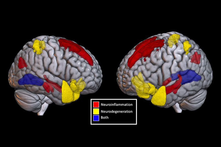
Neuroinflammation, which is defined by increased levels of inflammation-causing proteins in the brain, is a key feature of these conditions.
Previous studies suggest neuroinflammation can be detected before the appearance of other brain changes that are typically used to diagnose NDs. This means it could be a marker for earlier diagnosis. Unfortunately, many current diagnostic tools cannot detect signs of neuroinflammation — or they require invasive procedures.
A recent study from UHN’s Krembil Brain Institute suggests free-water diffusion (FWD) MRI could fill this diagnostic gap.
Free water is water in the brain that is not contained inside brain cells. FWD MRI measures how much of this water is present and how it moves. Changes in its amount, location, or movement can signal processes like neuroinflammation and cell death.
The Krembil Brain Institute team analyzed MRI data and blood samples from 367 patients with various NDs collected through the Ontario Neurodegenerative Research Initiative (ONDRI) database. They assessed FWD patterns in brain scans and measured blood levels of two protein markers — GFAP (linked to neuroinflammation) and NfL (linked to cell damage).
Using machine learning, the researchers found that specific FWD patterns could predict GFAP levels, suggesting the technique may detect neuroinflammation. They also identified differences in FWD patterns between different NDs — hinting that FWD MRI could be used to help identify not only the presence of neuroinflammation but also the type of ND in its early stages.
“Further studies in larger and more diverse groups are needed to confirm reliability and effectiveness,” says Dr. Carmela Tartaglia, senior author of the study, a Krembil clinician investigator, and a professor at the University of Toronto’s Tanz Centre for Neurodegenerative Diseases. “But we believe FWD MRI has great potential as a diagnostic tool.”
As a non-invasive technique that uses existing technologies, FWD MRI offers a way to make ND diagnosis at an earlier stage easier and more widely available.
For more patients, this could mean a better chance at effective management of and a life less impacted by neurodegeneration.
The first author of this study is Vishaal Sumra, a PhD candidate at the University of Toronto’s Institute of Medical Science.
This work was supported by generous donors to UHN Foundation.
No one ever changed the world on their own but when the bright minds at UHN work together with donors we can redefine the world of health care together.