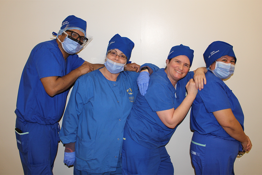
John Roeger, 3D technical specialist, vascular and interventional radiology technologist at the Joint Department of Medical Imaging, uses a stylus and special software to create vital 3D images. (Photo: UHN)
When medical radiation technologist John Roeger gets a call from a surgeon to create a 3D model of a brain aneurysm, time is of the essence.
A brain aneurysm is an abnormal bulge or balloon in the wall of a blood vessel that can cause internal bleeding and become life-threatening. John, a 3D technical specialist at the Joint Department of Medical Imaging (JDMI), starts by viewing a CT scan of the brain. Then, with a digital pen and special software, he “carves out” and isolates the aneurysm and creates several 3D, reconstructed images.
The images are then available to the surgeon.
“These images help our neurosurgeons evaluate the real size and shape of the aneurysm, look at the geometry and understand the relation of it to the surrounding blood vessels, so that they can make the best treatment decisions,” says John, who has worked for more than 30 years at JDMI, which covers UHN, Sinai Health System and Women’s College Hospital.
For the past two decades, John has worked in the 3D Lab at Toronto Western Hospital, where he says, “All the magic happens.”
According to Dr. Ronit Agid, neuroradiologist at UHN, 3D images are crucial to providing the most accurate diagnosis and best care possible.
“In some cases, an aneurysm can be so small that it’s missed on a regular scan,” Dr. Agid says. “But when you’re looking at a 3D image, the aneurysm is looking right at you and becomes a lot more obvious.
“It’s also critical for treatment planning. In a recent case, when I looked at the CT scan images it appeared to be a very straightforward aneurysm, but after I saw the 3D images, I realized that it wasn’t just one balloon, it was a double balloon and that changes the treatment plan completely.”
In 2005, its first year of operation, the 3D lab produced images for 50 cases. Now, John completes eight to 14 cases each day with speed and precision, supporting patients from UHN and Sinai Health.
John creates reconstructions that support everything from a life-saving liver transplant to shoulder surgery.
“Shoulder imaging is a big part of what we do too,” John says. “Orthopeadic surgeons are able to see the condition of the bone, the topography and determine if they can go in and do their surgery.”
‘Working together to increase clinical care and benefit patients’
Every week, John supports an average of 15 patients who require facial reconstruction surgery. This type of surgery can help correct abnormalities from birth defects, traumatic injury, disease or aging.
He also supports breast cancer survivors who are undergoing breast reconstruction surgery by providing precise images of the abdomen.
“The surgeons often take a section of the abdomen and use that tissue and native artery to reconstruct the breast,” John says.
Dr. Robert Bleakney is a staff musculoskeletal radiologist at the JDMI and has seen how 3D imaging continues to evolve and support his work.
“It used to be just bone that we can see, now we see tumours and soft tissues masses,” Dr. Bleakney says. “John is producing really sophisticated reconstructions that can really help us plan.
“Sometimes it’ll make the difference between limb preservation and amputation – so it helps inform very important decisions,” Dr. Bleakney says. “Sometimes John will go ahead on his own and complete different measurements and reformats which have been excellent.
“At the end of the day, we’re all working together to increase clinical care and benefit patients.”
The JDMI has ordered three state-of-the-art computers that will integrate with the latest software and ensure the 3D lab remains at the forefront of medical imaging practice. The department also plans to recruit and train current medical radiation & imaging technologists (MRITs) on staff to use the 3D technology.


