By Derek Malcolm
In late 2020, the Peter Munk Cardiac Centre unveiled the Dr. Susan Lenkei-Kerwin Catheterization Laboratory – also known as the Cath Lab – a new state-of-the-art facility at Toronto General Hospital that’s the first of its kind in Canada. It employs sophisticated technology, including the game-changing Philips Azurion platform, the first installation of the system in Canada, to treat a wide range of patients with complex heart disease, such as those with angina or congenital heart disease, with minimally invasive techniques. These procedures allow clinicians to get at the heart with tiny incisions, or no incision at all, instead of opening up the chest. The lab’s high-quality imaging equipment gives physicians a better look at the heart, which translates into safer and faster procedures for their patients. Not having to break any bones also increases a patient’s post-operation recovery time.
The Cath Lab includes two brand new procedure suites (with two more coming soon) and 28 new patient beds, all made possible thanks to donor support.
“I cannot overstate the significance of the Cath Lab redevelopment to the Peter Munk Cardiac Centre,” says Dr. Barry Rubin, Medical Director and Chair of the Peter Munk Cardiac Centre. “It’s through the generosity of our donors that this reimagined space will be used to treat patients with the most complex cases of heart and vascular disease.”
Here’s a look inside the Cath Lab and the advanced equipment teams use for better patient outcomes.
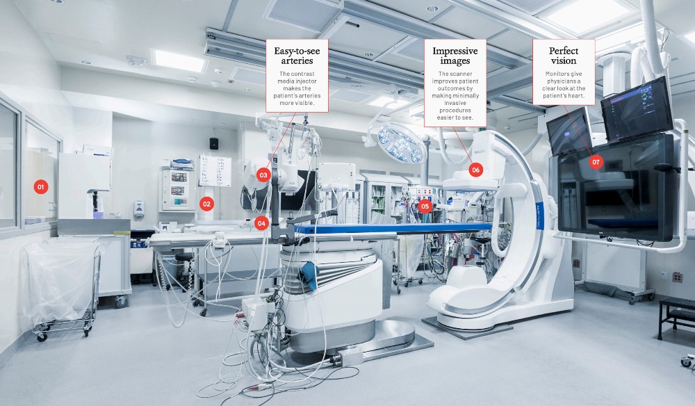
01 Control room
During a procedure, an X-ray technician or nurse is stationed behind glass in this separate room to monitor the patient’s heart rate, blood pressure, oxygen level and heart rhythm. That person, who is in constant communication with the team inside the lab, watches for any sudden changes in the patient. The scanner’s images are also shown on screens inside this room, so the technician can support the surgical team in real time.
02 Lead shield
Depending on the procedure, several nurses will circulate in and out of the room to assist. When not at the procedure table, they are stationed behind this lead shield to protect themselves
from radiation exposure.
03 Contrast media injector
It looks like a laser gun from an old sci-fi movie, but this machine is a power injector that shoots an iodine-based contrast media, or “X-ray dye,” into a patient’s arteries to make them more visible when photographed with the scanner. Thanks to the dye, cardiologists can clearly see the outlines of the arteries on an X-ray as they’re guiding catheters through the body.
04 Control panels
The interventional cardiologists – there are usually two – are positioned here. They work together to perform the procedure and control the position of the scanner to get the best images possible. The panels are positioned at groin level, as this is where the catheter and tubes are inserted.
05 Anesthetist’s station
Most of the procedures in the Cath Lab are done while the patient is awake but under sedation. Some longer and more complicated surgeries, including heart attack cases, require a general anesthetic. In both situations, the anesthetist is stationed here to monitor the patient.
06 Philips Azurion scanner
Cardiac catheterization is a term to describe procedures that help a cardiologist diagnose and treat heart problems, such as coronary artery disease or congenital heart defects. It’s minimally invasive because clinicians insert a small needle (about two millimetres) into the main artery or vein in the groin area and insert a thin tube called a catheter through that needle into the blood vessel. Tiny tools and devices are then inserted through the catheter and are guided to the heart to perform procedures like coronary angioplasties (in which small balloons or stents are inserted to open narrowed heart blood vessels) or to fix leaking or narrowed heart valves. To do these and other catheter-based procedures successfully, physicians need to clearly see what they’re doing. The better the picture, the better decisions they can make. The system uses two X-ray imaging scanners simultaneously, so cardiologists can move and position them over the patient on the table to view arteries and the cardiac system in real time, with unparalleled clarity and quality. The Azurion also uses significantly less radiation than older equipment, which reduces exposure to patients and staff.
07 Monitors
These three monitors are the eyes of the Azurion scanner. They display ultra-detailed live X-ray video images as physicians and technicians perform procedures on extra-large screens so everyone in the room can clearly see what is happening inside the patient’s blood vessels and heart. With their attention tuned to these screens, physicians can clearly see what they’re doing inside the patient in high resolution. The two monitors on top allow staff to view not only live images but also still ones they’ve taken as reference points to help them guide catheters through the arteries. One remarkable feature of the Azurion is that additional images, such as still X-rays and ultrasounds from previous tests, can be imported and overlaid, with the machine figuring out, in three dimensions, how the images correspond to one another. This system, called the EchoNavigator, helps interventional cardiologists see a 3D view of the heart.
This article originally appeared in the 2021 Peter Munk Cardiac Centre annual report. Read it here.
Photos by Tim Fraser.