By Elizabeth-Chorney Booth
When Eduard Savchuk went to see his family doctor with complaints of an increasingly hoarse voice last year, he had no inkling there was a massive tumour inside his chest. The 56-year-old electrician from Newmarket, Ont. had no pain, felt no unusual pressure and wasn’t experiencing any obvious organ failure. Like most of us, he – and, initially, his doctor – assumed his loss of voice was the result of a run-of-the-mill virus or a simple problem with his vocal cords that would pass on its own.
Not only did Savchuk’s voice not repair itself, but by the summer of 2019, he couldn’t speak at all and had to rely on his wife to make phone calls for him. After a return trip to his GP’s office, Savchuk was sent for diagnostic scans, which revealed a four-kilogram cancerous tumour entwined around his heart and airway. The tumour compressed the nerve to his voice box – an ominous sign.
“I didn’t know about the tumour at all,” Savchuk says. “I couldn’t understand how such a big mass could exist inside my body when I didn’t feel anything. It was so strange to me, to have what I had inside of me, wrapped around my heart.”
I was surprised when they gave me a model of my heart and my tumour. It let me see how the procedure would go. I was absolutely astonished.” Photo by Tim Fraser.
Making the impossible possible
Savchuk went through a round of chemotherapy, which didn’t shrink the tumour. His local oncology team decided it was too large and too close to his organs to remove safely. Thinking there was little to be done other than to keep Savchuk comfortable as the tumour slowly took his life, in a last-ditch effort, his doctors referred the case to Dr. Shaf Keshavjee, UHN’s Surgeon-in-Chief and the James Wallace McCutcheon Chair in Surgery, who is known for making seemingly impossible operations possible.
Even for a surgeon with Dr. Keshavjee’s vast experience, Savchuk’s tumour was challenging. Shaped sort of like a Mickey Mouse head, the tumour had “ears” that wrapped behind and pressed into the heart, and interfered with both his airway and his aorta. Dr. Keshavjee knew the tumour would be fatal if it went untreated, but he also didn’t want to go into surgery if he wasn’t confident the procedure would work. He could see the general placement of the tumour through CT and MRI scans, but they didn’t give him enough critical spatial information to determine if he could operate safely.
“It was already starting to obstruct his heart and his vena cava. So he was slowly dying,” recalls Dr. Keshavjee, explaining that while the tumour was growing slowly, the pressure was becoming more than Savchuk’s organs could withstand. “Even though he didn’t feel sick, there was this huge thing inside of him, literally the size of his head.”
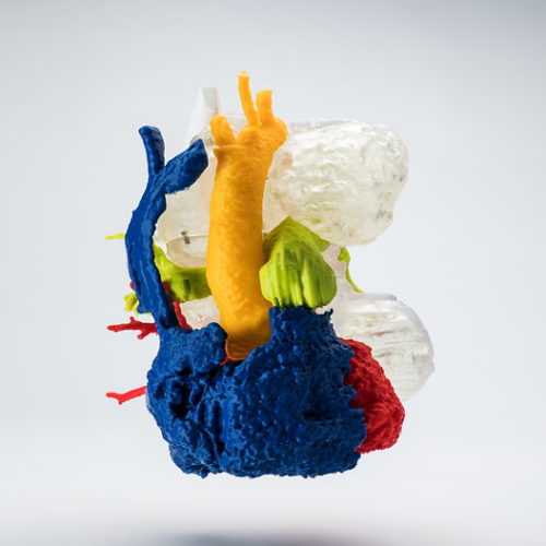
The 3D model of Shavchuk’s tumour that doctors used to prepare for surgery. Photo by Tim Fraser.
A team approach
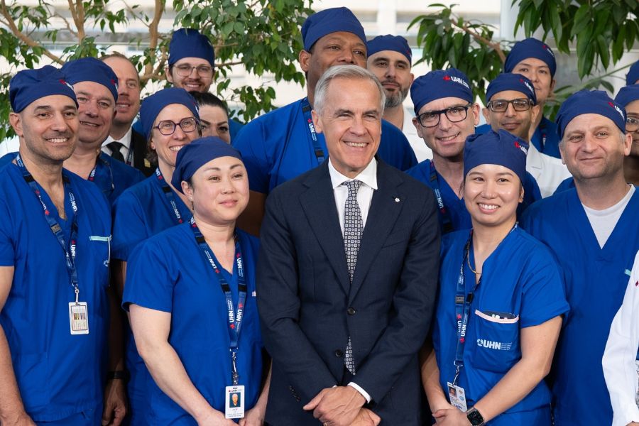
Because of his experience with similarly complicated tumours, Dr. Keshavjee thought he had a good shot of saving Savchuk, but he knew it wasn’t a one-surgeon job. Thankfully, in addition to attracting some of the highest-rated surgeons in Canada, the Sprott Department of Surgery has earned a reputation for its highly collaborative approach to patient care. Surgeons with expertise in treating one organ can call in equally skilled colleagues with expertise in another. The department’s multidisciplinary approach also extends to Sprott Surgery’s various teams of engineers, computer scientists and other healthcare-adjacent professionals. Safely getting Savchuk’s tumour out of his chest was going to take careful planning and teamwork.
Dr. Keshavjee started by reaching out to a colleague, cardiovascular surgeon Dr. RJ Cusimano. Dr. Keshavjee knew that in addition to years of experience dealing with cardiovascular tumours, Dr. Cusimano, who holds the David & Stacey Cynamon Professorship in Cardiovascular Surgery Innovation and Education, and is also part of the Peter Munk Cardiac Centre’s team, was open to using technology to come up with outside-the-box surgical solutions.
Drs. Keshavjee and Cusimano next tapped into the expertise of Dr. Azad Mashari, an anesthesiologist in the Department of Anesthesia and Pain Management at Toronto General Hospital, who has the ability to create 3D printed models derived from patients’ diagnostic scans. The models gave the surgeons a more accurate picture of how Savchuk’s tumour was interacting with his heart so they could decide whether surgery was even a possibility. More importantly, the surgical team could then simulate where specific incisions might be made to extricate the tumour from the middle of Savchuk’s chest.
“With the 3D model, we could figure out how to dismantle the tumour like a jigsaw puzzle before we even went into operation.” Dr. Shaf Keshavjee. Photo by Tim Fraser.
Holding his heart in his hands
3D modelling may not seem like the natural purview of an anesthesiologist, but just as Sprott Surgery employs some of the world’s best surgeons, its anesthesiologists are also world-class.
It’s not uncommon for a high-level anesthesiologist like Dr. Mashari to perform transesophageal echocardiograms on patients, which involve an internal ultrasound that gives surgeons a better look at a patient’s heart immediately before surgery. Dr. Mashari had already been using internal ultrasounds to create 3D ultrasound images (much like the videos expectant parents get with prenatal ultrasounds), which led to the idea to create 3D models from CT scans that surgeons could look at well before surgery.
“The aspect of making 3D models is not routinely part of anesthesia,” says Dr. Mashari. “It’s part of the innovative mindset that exists at UHN.”
Dr. Mashari and his colleague Joshua Qua Hiansen spent more than 15 hours in the lab using information from a series of Savchuk’s CT scans to develop the life-size 3D model, which took 40 hours to produce on a 3D printer. The result was a colour-coded model that not only showed Drs. Keshavjee and Cusimano exactly how the tumour was placed, but for the first time also gave Savchuk a clear visual representation of what was going on in his body. Before surgery, Savchuk got to hold the model of his tumour and heart, which somewhat quelled his fears, because he was able to understand exactly what his doctors were going to do.
“I’m a technical guy – I work with electronics,” Savchuk says. “I like to understand my body like I would a computer. The model helped me understand those parts of my body and how complicated they can be. After seeing it, and hearing Drs. Keshavjee and Cusimano’s explanation, it was more clear to me what would happen on the surgical table.”
“You can see how intertwined the tumour is with his heart. The patient didn’t know how complex it was until he held the model in his hands.” Dr. RJ Cusimano. Photo by Tim Fraser.
Smart OR for smarter surgery
Dr. Mashari’s model wasn’t the only UHN-specific technical element that helped the Sprott surgeons successfully remove Savchuk’s tumour. The 3D images of Savchuk’s heart and tumour – both the physical model and the digital representation that could be manipulated on a computer screen – were important for planning before the surgery, but the surgeons also needed to see the images during surgery. While computer displays are not uncommon in operating rooms, to access them, surgeons typically have to scrub in and scrub out, wasting precious time while the patient’s chest is open.
Bringing technology into the surgical process requires support so that it doesn’t unintentionally impede surgeons as they concentrate on more delicate work. To address this, UHN has developed a program dubbed “Surgility,” which incorporates the expertise of on-site engineers in operating rooms. In Savchuk’s case, the surgeons worked with Jimmy Qiu, manager of engineering at the Techna Institute for the Advancement of Technology for Health at UHN, to set up displays on moveable arms adjacent to the operating table – similar to what would be used for laparoscopic surgery – so that the surgeons could quickly access the 3D imaging as they worked. Qiu says the direct relationship between his engineering team and Sprott Surgery makes it easier to come up with innovative solutions for these complex cases.
“The patient knew that if he did nothing, the tumour would kill him. Exactly how long it would take, we didn’t know.” Dr. Azad Mashari. Photo by Tim Fraser.
“Traditionally in an academic hospital setting, research engineers work in a lab and have a very siloed experience,” Qiu explains. “We typically don’t see the pain points and have that interaction with the stakeholders. When engineers are embedded in the OR, that leads to more engagement and interaction, and the surgeons are more open in their real-time communication as well. We all learn more.”
All of these pieces coming together – the engineering, the 3D imaging and printing, and the collaboration between surgeons, anesthetists, perfusionists and nurses – is what got that tumour out of Savchuk’s chest. The extraction surgery was performed in December 2019. Dr. Mashari was the anesthesiologist, while Drs. Keshavjee and Cusimano successfully removed the tumour within the span of four or five hours. Dr. Keshavjee estimates it could have taken up to 10 hours without the opportunity to plan in advance with the aid of the 3D model, which would have put Savchuk at additional risk. While it would be overly simplistic to say the procedure was “routine,” the advance planning and ability to reference the model meant there was an efficiently pre-developed operative plan and few surprises or complications for the surgeons.
Savchuk’s speaking voice is still husky, but he’s now able to talk with ease and has returned to work. His doctors are confident the tumour was completely removed, and Savchuk’s most recent round of tests confirm he is cancer-free.
While the outcome is what matters most to Savchuk and his loved ones, the success of the operation is also significant to Drs. Keshavjee and Cusimano, in that it illustrates what’s possible when high-performance surgical teams work together and leverage the breadth of talent and expertise of all the medical professionals at UHN. It’s this approach that makes Sprott Surgery capable of taking such challenging cases and giving real hope to patients who thought they’d run out of options.
“We don’t say no to people here. We say, ‘We’ll find out,’” Dr. Cusimano says. “If (Savchuk) had come to us three years earlier, we may not have been able to help him. But every year we build a little bit more. Who knows what’s going to happen next? It’s a constant evolution.”
“It’s important to appreciate the spatial context of the tumour. Dr. Keshavjee wanted to visualize the data on the monitor as the surgery was happening.” Jimmy Qiu. Photo by Tim Fraser.
This article was featured in the 2020 Sprott Department of Surgery magazine, view the whole magazine online here!
Photos by Tim Fraser.